Oncolytic Peptides are rapidly emerging as one of the most exciting innovations in cancer immunotherapy. These engineered peptide molecules do more than simply kill tumor cells. They also activate the immune system and reshape the tumor microenvironment. As a result, researchers are studying Oncolytic Peptides as both direct cytotoxic agents and immune modulators.
Unlike traditional chemotherapy, which broadly targets dividing cells, Oncolytic Peptides are designed to exploit structural differences in cancer cell membranes. At the same time, they trigger immunogenic cell death, which transforms tumors into visible immune targets. Therefore, this dual mechanism has positioned Oncolytic Peptides at the forefront of translational oncology research.
Let us break down how these molecular disruptors work and why the field is gaining momentum.
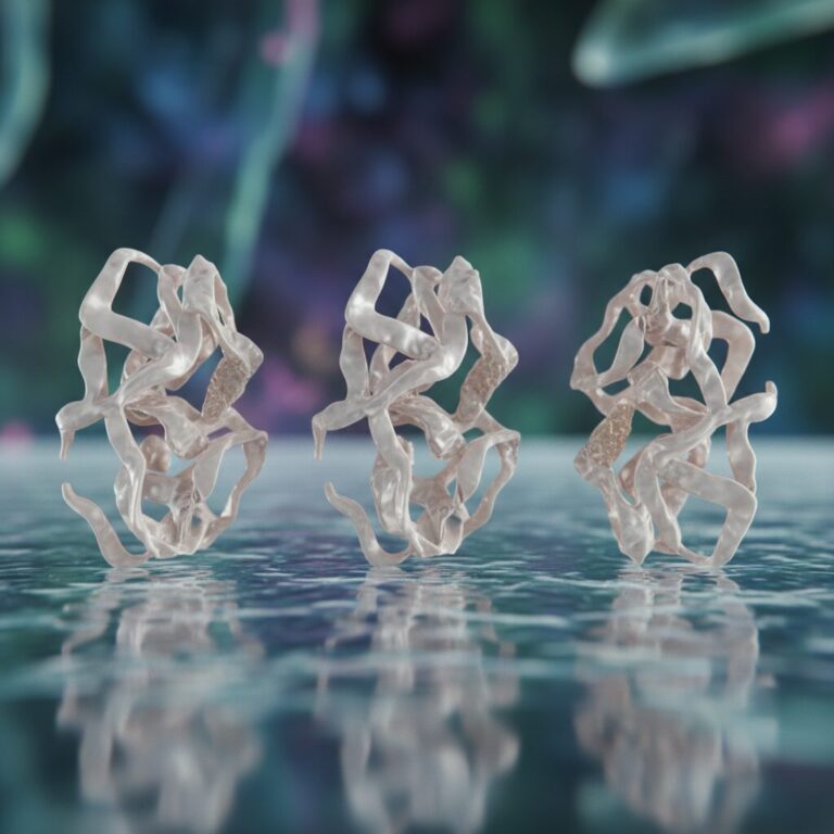
Oncolytic Peptides are short synthetic or engineered amino acid sequences designed to selectively destroy malignant cells. Many are inspired by antimicrobial host defense peptides such as defensins and cathelicidins. These natural peptides evolved to target microbial membranes. Scientists adapted their structure to preferentially target cancer cells.
Cancer cell membranes differ from healthy cells in several ways. For example, tumor membranes often expose negatively charged phospholipids like phosphatidylserine on their outer surface. In contrast, healthy cells typically keep these lipids on the inner leaflet of the membrane.
Because many Oncolytic Peptides carry a positive charge, they are electrostatically attracted to tumor membranes. Additionally, most are amphipathic, meaning they contain both hydrophilic and hydrophobic regions. This structure allows them to insert into lipid bilayers efficiently.

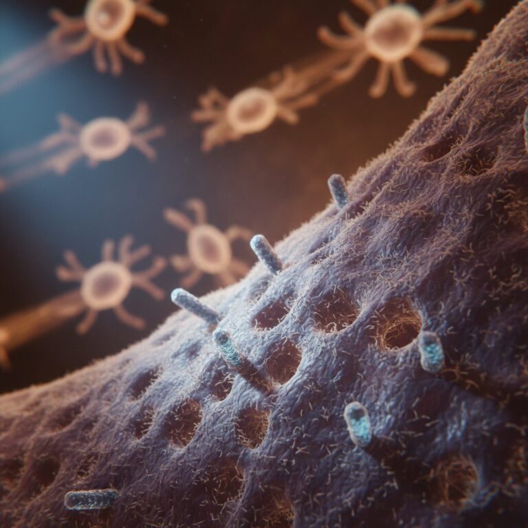
The first function of Oncolytic Peptides is membrane disruption. After binding to the cancer cell surface, the peptide inserts into the membrane. Subsequently, it forms pores or destabilizes the lipid bilayer.
As a result, the membrane loses integrity. Cellular contents leak out, osmotic balance collapses, and the tumor cell undergoes lysis. This process is typically rapid and does not rely on traditional apoptosis pathways.
One extensively studied candidate is LTX-315, a synthetic peptide derived from bovine lactoferricin. LTX-315 not only disrupts membranes but can also penetrate into tumor cells. Inside the cell, it destabilizes mitochondria.
Since mitochondria regulate energy production and apoptosis signaling, mitochondrial damage amplifies tumor cell death. Consequently, Oncolytic Peptides such as LTX-315 demonstrate multi-layered cytotoxic effects.
Importantly, this destruction is not silent.
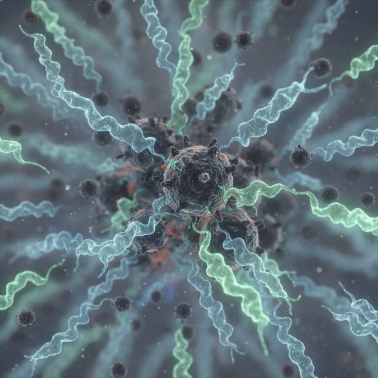
When tumor cells die through traditional apoptosis, they often evade immune detection. However, Oncolytic Peptides frequently induce immunogenic cell death. This form of cell death releases danger signals known as danger associated molecular patterns or DAMPs.
These molecules alert the immune system that something abnormal has occurred.
Key DAMPs released after treatment with Oncolytic Peptides include:
Because of these signals, dendritic cells engulf tumor debris and process tumor antigens. Subsequently, they present these antigens to CD8 positive cytotoxic T lymphocytes. These T cells then seek out and destroy remaining cancer cells.
Therefore, Oncolytic Peptides convert tumors from immune cold environments into inflamed, immune active sites.
Beyond DAMP release, Oncolytic Peptides may activate innate immune pathways. For example, mitochondrial disruption can release mitochondrial DNA into the cytoplasm. This DNA activates the cGAS STING signaling pathway.
Activation of cGAS STING leads to the production of type I interferons. These interferons enhance dendritic cell maturation and T cell priming. As a result, the immune response becomes more robust and sustained.
Preclinical studies also suggest involvement of Toll like receptor pathways and MyD88 dependent signaling. However, these mechanisms are still being clarified in ongoing research.
Taken together, Oncolytic Peptides function as both tumor disruptors and immune amplifiers.
Oncolytic Peptides have entered early phase clinical trials. LTX-315 has been evaluated in patients with melanoma, sarcoma, and other solid tumors. These studies primarily used intratumoral injection.
Reported findings from Phase I and Phase II studies include:
In some basal cell carcinoma trials, high objective response rates were reported. Larger confirmatory studies are ongoing. Readers can review active trials on ClinicalTrials.gov by searching LTX-315.
Other investigational Oncolytic Peptides include CyPep-1, EP-100, and derivatives of LL-37. Each is being evaluated for safety, tumor selectivity, and immune activation.
Checkpoint inhibitors such as anti PD 1 therapies have transformed oncology. However, they work best in tumors already infiltrated by T cells.
Oncolytic Peptides may help generate that infiltration. By inducing immunogenic cell death and releasing tumor antigens, they prime the immune system.
Therefore, combination trials are testing Oncolytic Peptides alongside checkpoint inhibitors. The strategy is straightforward. First, Oncolytic Peptides disrupt the tumor and activate immune signaling. Next, checkpoint inhibitors prevent immune exhaustion.
Together, these approaches may enhance durable anti tumor responses.
Although Oncolytic Peptides show promise, delivery remains a challenge. Most clinical studies rely on direct tumor injection. While effective for accessible tumors, this method limits use in deep or metastatic disease.
Systemic administration presents additional obstacles. Peptides can be degraded by circulating proteases. They may also clear rapidly from the bloodstream.
To address this issue, researchers are developing nanocarrier systems. These include liposomes, polymer nanoparticles, and metal organic frameworks.
Such carriers can protect Oncolytic Peptides from degradation and enhance tumor specific delivery. Furthermore, some systems are designed to release the peptide in response to acidic tumor environments.
Tumors evolve. Over time, they may alter membrane lipid composition. They may also activate survival signaling pathways that counteract peptide induced stress.
Consequently, understanding resistance mechanisms is critical for optimizing Oncolytic Peptides.
Researchers are investigating lipidomic and glycomic profiling to predict tumor susceptibility. Biomarker development will likely guide patient selection and improve clinical outcomes.
Oncolytic Peptides differ from oncolytic viruses because they do not replicate. This simplifies certain safety concerns. However, they remain classified as investigational immunotherapeutics.
Manufacturing consistency, stability testing, and pharmacokinetics must meet regulatory standards. Additionally, long term safety data will be essential.
As the field matures, standardized frameworks for peptide immunotherapy evaluation will continue to evolve.
Cancer immunotherapy has shifted from broad cytotoxic approaches toward precision immune modulation. Oncolytic Peptides align with this trend.
They combine:
Because of this multi dimensional activity, Oncolytic Peptides represent a hybrid strategy that bridges cytotoxic therapy and immunotherapy.
Although the field remains early stage, translational momentum continues to grow. With improvements in delivery systems, biomarker identification, and combination therapy design, Oncolytic Peptides may become an important component of future cancer treatment protocols.
Oncolytic Peptides are reshaping how researchers think about cancer therapy. Rather than choosing between direct tumor destruction and immune activation, these engineered molecules accomplish both.
While challenges remain in systemic delivery and resistance, ongoing clinical trials and technological advances are steadily moving the field forward.
As research expands, Oncolytic Peptides may help convert immune silent tumors into responsive targets. For oncology innovation watchers, this is a space worth following closely.
¹ Kepp, O., Deng, X., Xue, E., Sveinbjørnsson, B., Rekdal, Ø., Galluzzi, L., & Kroemer, G. (2026). Immunobiological mechanisms of action of oncolytic peptides. Journal for ImmunoTherapy of Cancer, 14(2), e013337. https://jitc.bmj.com/content/14/2/e013337
² Koo, D. J., Sut, T. N., & Tan, S. W. (2022). Biophysical Characterization of LTX-315 Anticancer Peptide Interactions with Model Membrane Platforms: Effect of Membrane Surface Charge. International Journal of Molecular Sciences, 23(18), 10558. https://pubmed.ncbi.nlm.nih.gov/36142646/
³ Eike, L. M., Yang, N., Rekdal, Ø., & Sveinbjørnsson, B. (2015). The oncolytic peptide LTX-315 induces cell death and DAMP release by mitochondria distortion in human melanoma cells. Oncotarget, 6(33), 34910–34923. https://pubmed.ncbi.nlm.nih.gov/26472184/
⁴ Zhou, H., Forveille, S., Sauvat, A., Yamazaki, T., Senovilla, L., Ma, Y., & Kroemer, G. (2016). The oncolytic peptide LTX-315 triggers immunogenic cell death. Cell Death & Disease, 7(3), e2134. https://pubmed.ncbi.nlm.nih.gov/26962684/
⁵ Li, X., Su, N., Yu, H., Yuan, X., Du, S., Zeng, C., He, C., Peng, L., Guo, H., Shi, F., Wang, Z., & Chen, Y. (2024). Hainanenin-1, an oncolytic peptide, triggers immunogenic cell death via STING activation in triple-negative breast cancer. Cell Communication and Signaling: CCS, 22(1), 352. https://pubmed.ncbi.nlm.nih.gov/39109040/
⁶ Li, X.-Q., Yamazaki, T., He, T., Alam, M. M., Liu, J., Trivett, A. L., Sveinbjørnsson, B., Rekdal, Ø., Galluzzi, L., Oppenheim, J. J., & Yang, D. (2024). LTX-315 triggers anticancer immunity by inducing MyD88-dependent maturation of dendritic cells. Frontiers in Immunology, 15, 1332922. https://pubmed.ncbi.nlm.nih.gov/38496155/
⁷ Sveinbjørnsson, B., Camilio, K. A., Haug, B. E., & Rekdal, O. (2017). LTX-315: A First-In-Class Oncolytic Peptide that Reprograms the Tumor Microenvironment. Future Medicinal Chemistry, 9(12), 1339–1344. https://pubmed.ncbi.nlm.nih.gov/28490192/
⁸ Vitale, I., Yamazaki, T., Wennerberg, E., Sveinbjornsson, B., Rekdal, O., Demaria, S., & Galluzzi, L. (2021). Targeting Cancer Heterogeneity with Immune Responses Driven by Oncolytic Peptides. Trends in Cancer, 7(6), 557–572. https://pubmed.ncbi.nlm.nih.gov/33446447/
⁹ Kantor, J., Bhatia, N., Green, L., & Jaffe, B. (2025). Calculated Objective Response Rate (ORR) of 97% from Post-Hoc Analysis of a Phase 2 Multicenter Study to Evaluate the Efficacy of VP-315, an Investigational Therapy for Basal Cell Carcinoma (BCC). Journal of Skin, 9(Supplement 1), S565. (NCT05188729)
¹⁰ Spicer, J., Marabelle, A., Baurain, J.-F., Jebsen, N. L., Jossang, D. E., Awada, A., Haugland, H. K., Rekdal, Ø., Linder, D., Ghert, M., Hjelle, T., Melin, S. A., & Strand, A. (2021). Safety, Antitumor Activity, and T-cell Responses in a Dose-Ranging Phase I Trial of the Oncolytic Peptide LTX-315 in Patients with Solid Tumors. Clinical Cancer Research, 27(10), 2755–2763. https://pubmed.ncbi.nlm.nih.gov/33542073/ (NCT01058616; NCT01986426)
¹¹ Al Musaimi, O., Armstrong, A., Coburn, F., & Nsereko, Y. (2025). Innovative Peptide Therapeutics in the Pipeline: Transforming Cancer Detection and Treatment. International Journal of Molecular Sciences, 26(14), 6815. https://pubmed.ncbi.nlm.nih.gov/40725089/ (General overview of peptide therapeutics in pipeline including clinical trial IDs for CyPep-1, EP-100, LL-37)
¹² Al Musaimi, O., Lombardi, L., Williams, D. R., & Albericio, F. (2022). Strategies for Improving Peptide Stability and Delivery. Pharmaceuticals, 15(10), 1283. https://pubmed.ncbi.nlm.nih.gov/36297395/
¹³ Xia, Y., Wei, J., Zhao, S., Guo, B., Meng, F., Klumperman, B., Schiffelers, R. M., & Yang, D. (2021). Systemic administration of polymersomal oncolytic peptide LTX-315 combining with CpG adjuvant and anti-PD-1 antibody boosts immunotherapy of melanoma. Journal of Controlled Release: Official Journal of the Controlled Release Society, 336, 262–273. https://pubmed.ncbi.nlm.nih.gov/34174350/
¹⁴ Ishikawa, K., Medina, S. H., Schneider, J. P., & Pitter, K. L. (2017). Glycan Alteration Imparts Cellular Resistance to a Membrane-Lytic Anticancer Peptide. Cell Chemical Biology, 24(2), 149–158. https://pubmed.ncbi.nlm.nih.gov/28162985/
All human research MUST be overseen by a medical professional.